A Bone Graft for Implants Rebuilds the Foundation for a Strong Smile
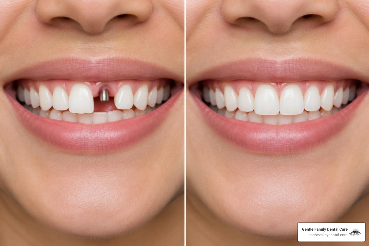
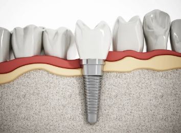
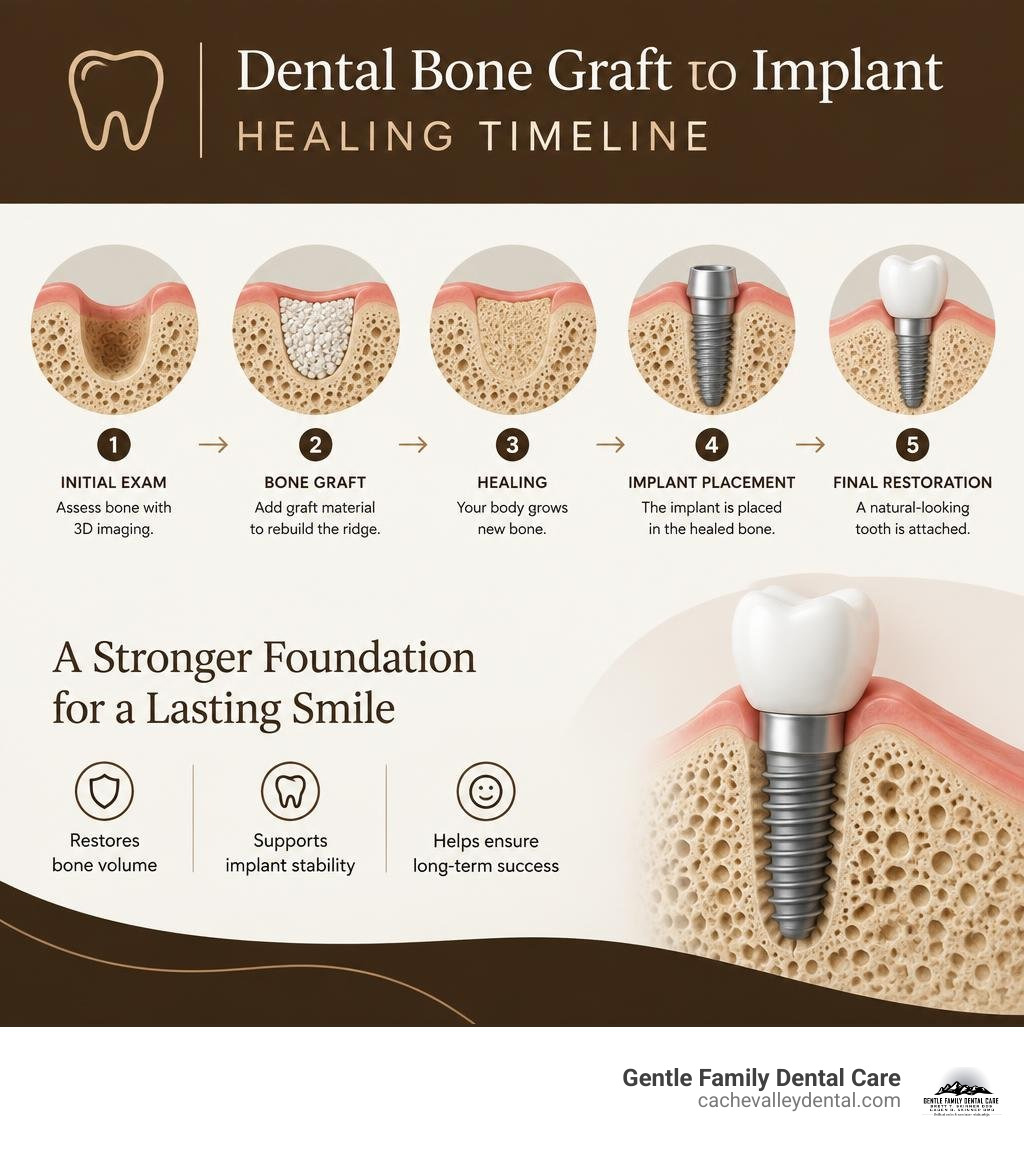
A bone graft for implants rebuilds jawbone that is too thin or too short to hold an implant securely. Your dentist uses 3D imaging to measure the bone at the planned implant site, selects the right graft approach, and allows the area to heal before placing the implant when needed.
Bone loss is common after an extraction because the jaw no longer receives the chewing forces that help maintain its shape. Gum disease, injury, and long-standing missing teeth can also reduce bone volume. A graft provides a framework where your body can grow and remodel new bone, helping create a stable base for an implant.
At Gentle Family Dental Care, we help patients in Logan and across Cache Valley, including North Logan, Smithfield, and Hyrum, understand their options clearly. A personalized exam and digital imaging can show whether grafting, a short implant, or another approach makes the most sense for your smile, comfort, timeline, and budget.
Why You Might Need a Bone Graft for Implants
Securing a long-lasting dental implant requires a solid bedrock of dense, healthy bone. Much like building a home on a stable foundation rather than shifting sand, placing an artificial root calls for adequate bone height, width, and mineral density. When a dental implant is positioned into the jaw, it relies on initial mechanical grip—known clinically as primary stability—to remain completely still while your living bone cells weave around it.
Without sufficient alveolar ridge volume, implants cannot achieve the required contact with host bone. Peer-reviewed literature in bone grafting techniques and materials emphasizes that establishing at least 1.5 to 2 millimeters of healthy bone thickness around an implant fixture is necessary to avoid future soft tissue recession and bone loss. When natural bone dimensions fall below these parameters, ridge augmentation restores the structural support required for predictable, long-term success.
Causes of Alveolar Bone Loss
The alveolar bone exists specifically to hold your natural teeth. Once a tooth is removed, that localized bone loses the mechanical stimulation generated by daily biting and chewing. Without these functional forces, your body begins resorbing the unneeded bone tissue. In fact, clinical studies demonstrate that alveolar ridges can lose 40% to 60% of their horizontal and vertical dimensions within the first two years following an extraction, with up to 25% of ridge width diminishing in the first twelve months alone.
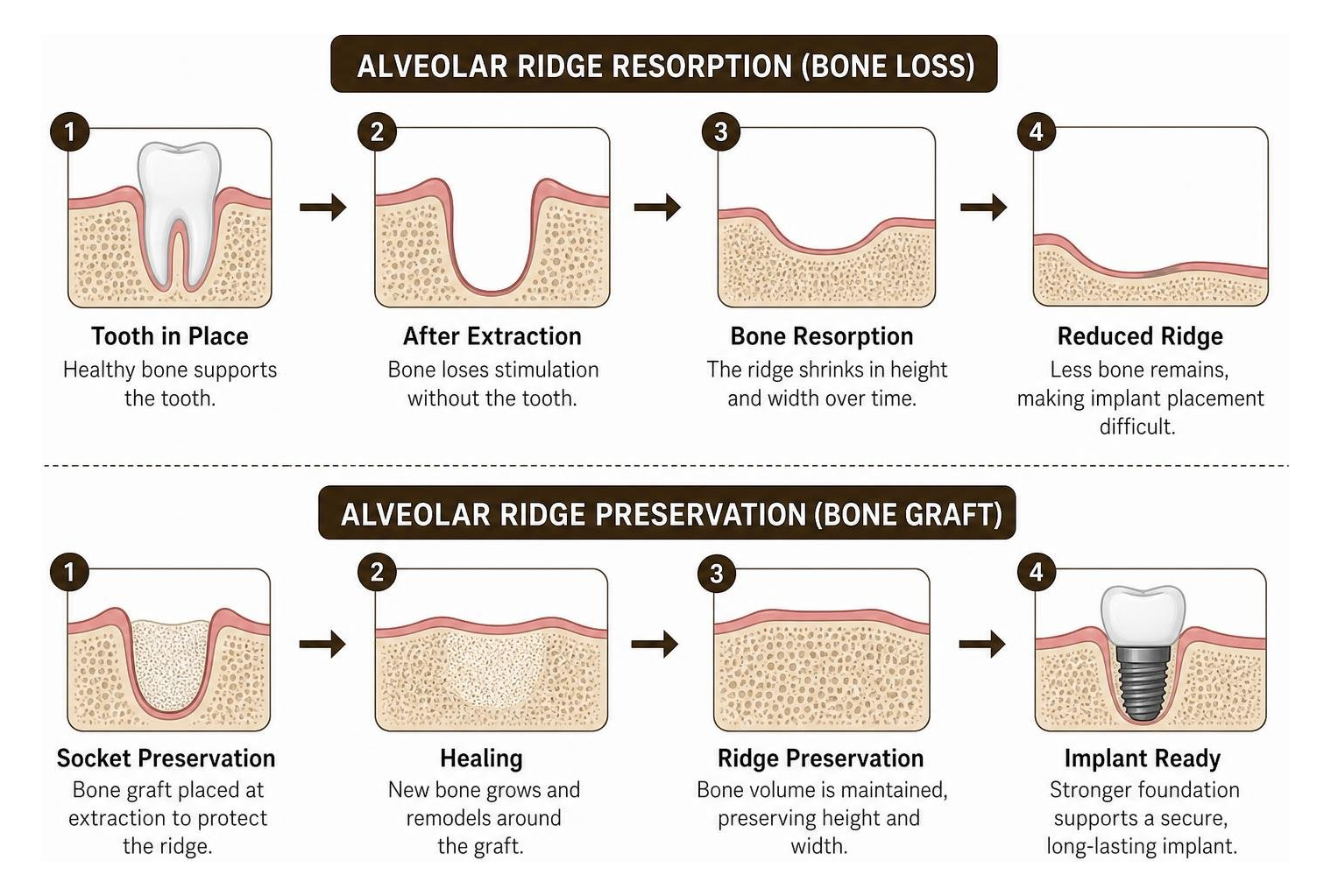
Beyond simple extractions, several underlying factors lead to bone loss:
- Periodontal Disease: Advanced gum infections destroy the periodontal ligament and the surrounding alveolar bone housing.
- Long-Term Tooth Loss: Leaving an empty space unaddressed for years allows progressive bone atrophy to flatten the ridge.
- Denture Pressure: Traditional removable appliances rest directly on the gums, causing continuous friction and accelerated ridge shrinkage over time.
- Trauma and Injury: Physical impacts or facial fractures can shatter delicate socket walls, compromising natural anatomy.
Evaluating Bone Quality and Quantity with 3D Imaging
To determine whether an augmented foundation is needed, our team at Gentle Family Dental Care utilizes low-radiation Cone Beam Computed Tomography (CBCT) scanning. Unlike standard two-dimensional x-rays that flatten anatomical structures, 3D imaging captures cross-sectional slices of your upper and lower jaws with sub-millimeter precision.
CBCT scans measure localized bone density in Hounsfield Units (HU) and map critical anatomical landmarks, such as the inferior alveolar nerve canal in the lower jaw and the maxillary sinus cavities in the upper jaw. High-resolution imaging reveals important anatomical differences: the mandible features dense cortical bone that provides substantial initial stability, whereas the posterior maxilla is naturally more porous and cancellous. Advanced research using 3D micro-CT assessment shows that mandibular sites often yield roughly 12.8% higher total bone volume during regeneration compared to maxillary sites, giving our clinicians essential diagnostic data to tailor your treatment plan right here in Logan.
Common Materials and Techniques for Ridge Augmentation
Bone grafting works through three primary biological mechanisms:
- Osteoconduction: The graft serves as a physical scaffold, allowing native capillaries and osteoblasts (bone-forming cells) to migrate across the defect.
- Osteoinduction: Chemical signals and growth factors actively recruit stem cells to differentiate into mature bone-forming cells.
- Osteogenesis: Living cells contained within the graft material directly produce new woven bone.
To successfully rebuild missing jawbone, grafting procedures maintain space underneath the gum tissue, allowing slow-growing bone cells to proliferate without being overtaken by rapidly dividing soft-tissue cells.

| Graft Material Type | Source Origin | Primary Biological Mechanism | Typical Resorption Rate | Best Clinical Application |
|---|---|---|---|---|
| Autogenous Bone | Patient’s own body (mandible, chin) | Osteogenic, Osteoinductive, Osteoconductive | Rapid to Moderate | Extensive vertical defects, complex reconstructions |
| Allograft | Human donor tissue (screened cadaveric) | Osteoconductive, mildly Osteoinductive | Moderate | Socket preservation, horizontal ridge augmentation |
| Xenograft | Bovine (cow) or Porcine (pig) mineral | Osteoconductive scaffold | Very Slow (long-term space maintenance) | Sinus lifts, buccal gap filling, contour preservation |
| Porcine Dentin Graft | Processed porcine tooth matrix | Osteoconductive, bioactive matrix | Balanced External Replacement | Socket preservation, highly cellular defect repair |
| Alloplast | Synthetic minerals (beta-tricalcium phosphate, biphasic ceramic) | Purely Osteoconductive | Variable based on chemistry | Contained defects, secondary cleft repairs |
Types of Bone Graft for Implants
Depending on the geometry and size of your bone defect, various grafting materials can be selected:
- Particulate Bone Grafts: Small granules of mineralized bone packed gently into contained defects or extraction sockets. They provide a high surface area for rapid revascularization.
- Block Grafts: Solid sections of cortical bone secured with micro-screws to correct severe horizontal or vertical ridge deficiencies larger than 5 to 6 millimeters.
- Porcine Dentin-Derived Grafts: Recent randomized clinical trials on porcine dentin-derived bone graft materials highlight that processed dentin retains natural collagen proteins and regular tubular pathways (1 to 2 micrometers in diameter). This architectural matrix delivers exceptional bone-graft integration (reaching upwards of 85%) and robust new bone formation compared to traditional bone xenografts.
- Engineered Scaffolds: Advanced biomaterials, including calcium-phosphate coated melt electrowritten polymeric scaffolds with gradient pore designs, continue to show excellent potential for guiding cellular infiltration and targeted vascularization (Abbasi et al., 2020).
Guided Bone Regeneration and Barrier Membranes
Soft tissue cells heal much faster than hard tissue cells. If left unprotected, the gums would quickly fill an extraction socket, preventing dense bone from forming. Guided Bone Regeneration (GBR) solves this problem by placing a protective barrier membrane over the grafted material.
Published insights in bone grafting for implant surgery highlight the critical role of these barriers:
- Resorbable Collagen Membranes: Derived from purified porcine or bovine collagen, these naturally break down over several months, eliminating the need for a second surgical procedure to remove them while maintaining cellular exclusion.
- CAD/CAM Titanium Meshes and Non-Resorbable Barriers: For large vertical augmentations, patient-specific 3D titanium meshes provide rigid mechanical protection. Clinical research shows that while titanium mesh alone performs with statistical non-inferiority to mesh covered with collagen sheets (producing roughly 29.8% to 30.8% newly mineralized bone volume), adding a membrane provides an extra protective buffer against early oral exposure.
Step-by-Step Surgical and Healing Timeline
Undergoing bone augmentation is a routine outpatient procedure designed to maintain comfort while restoring functional bone volume. Integrating this therapy alongside our general dentistry services ensures seamless oral health management from your initial exam to final restoration.
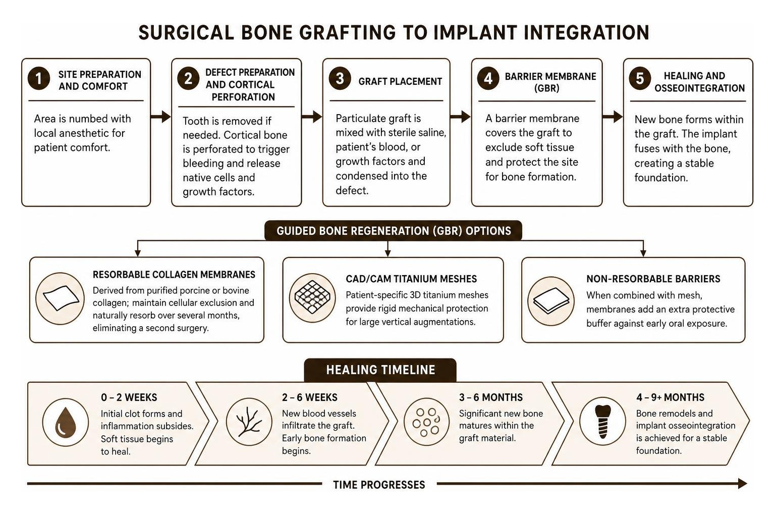
What to Expect During a Bone Graft for Implants Procedure
- Site Preparation and Comfort: The target area is completely numbed using modern local anesthetics. We prioritize a gentle, relaxing environment throughout your appointment.
- Defect Preparation and Cortical Perforation: If a damaged tooth is present, it is carefully removed to protect delicate socket walls. Tiny perforations are made into the dense outer cortical bone to encourage bleeding, releasing native osteogenic cells and growth factors into the site.
- Graft Placement: Particulate graft material is mixed with sterile saline, the patient’s own blood, or concentrated growth factors, and condensed smoothly into the bone deficiency.
- Membrane Adaptation and Closure: A resorbable collagen barrier is trimmed to cover the graft boundaries, protecting the graft particles. The gum tissue is repositioned and secured with precise, tension-free sutures.
Osseointegration and Recovery Milestones
Healing follows an organized biological trajectory:
- Weeks 1 to 2 (Early Soft Tissue Closure): Gums knit closed over the surgical site. Mild swelling and tenderness resolve within a few days.
- Months 1 to 3 (Osteoconduction and Mineralization): New blood vessels penetrate the scaffold. Osteoblasts deposit immature, low-mineralized woven bone across the graft granules.
- Months 3 to 6 (Remodeling into Mature Bone): The body replaces woven bone with load-bearing, high-mineralized lamellar bone. Micro-CT studies demonstrate that by 6 months, mature bone tissue accounts for nearly 30% of total site volume, with residual graft particles stably integrated into the surrounding matrix.
- Implant Placement: Once the site achieves solid clinical density, your dental implant is placed. Clinicians look for an insertion torque threshold above 30 to 35 Ncm, confirming excellent primary stability for long-term chewing strength.
Frequently Asked Questions About Dental Bone Grafting
If you have additional questions regarding your treatment, explore our patient dental faqs or discuss them directly with our clinical team.
Does a dental bone graft procedure hurt?
Most patients find that getting a bone graft involves minimal discomfort. The procedure is performed under local anesthesia, meaning you will feel gentle pressure during placement but no sharp sensation. Following surgery, any mild aching is typically managed with over-the-counter pain relievers such as ibuprofen or acetaminophen for two to three days.
How long after a bone graft can I receive an implant?
Healing timelines vary based on defect size and anatomical location. For standard extraction socket preservation using particulate bone, the site is typically ready for an implant in 3 to 4 months in the dense lower jaw, and 4 to 6 months in the upper jaw. Larger block grafts or extensive sinus floor elevations may require 6 months to mature into high-density bone.
Can dental implants be placed without grafting in thin bone?
In select anatomical scenarios, alternatives may avoid extensive bone grafting:
- Short Implants: When vertical bone height is limited in the posterior lower jaw, short implants (7 mm or shorter) can achieve clinical survival rates exceeding 90% while significantly reducing surgical complexity.
- Ridge Splitting: If the ridge has adequate vertical height but lacks horizontal thickness, mechanical ridge expansion allows simultaneous implant placement by gently widening the existing bone walls.
- Tilted Implants: Strategically angling implants allows clinicians to engage denser regional bone while bypassing anatomic structures like the maxillary sinus.
Restoring Your Smile Foundation in Cache Valley
At Gentle Family Dental Care, we take pride in delivering compassionate, state-of-the-art restorative and implant dentistry for families throughout Logan, North Logan, Smithfield, and Hyrum. With more than two decades of clinical experience, our bilingual team provides personalized, gentle care tailored to your exact oral health needs.
Whether you need a single-tooth socket preservation or comprehensive ridge augmentation, we are dedicated to helping you achieve a functional, lasting smile right here in the heart of Cache Valley. Contact our office today to schedule an implant consultation and take the first step toward restoring your oral health.
References
Abbasi N, Lee RSB, Ivanovski S, Love RM, Hamlet S. “In vivo bone regeneration assessment of offset and gradient melt electrowritten (MEW) PCL scaffolds..” Biomaterials research, 2020. PMCID PMC7529514.